Anatomy & Physiology
Typical Vertebra (Superior & Lateral Views)
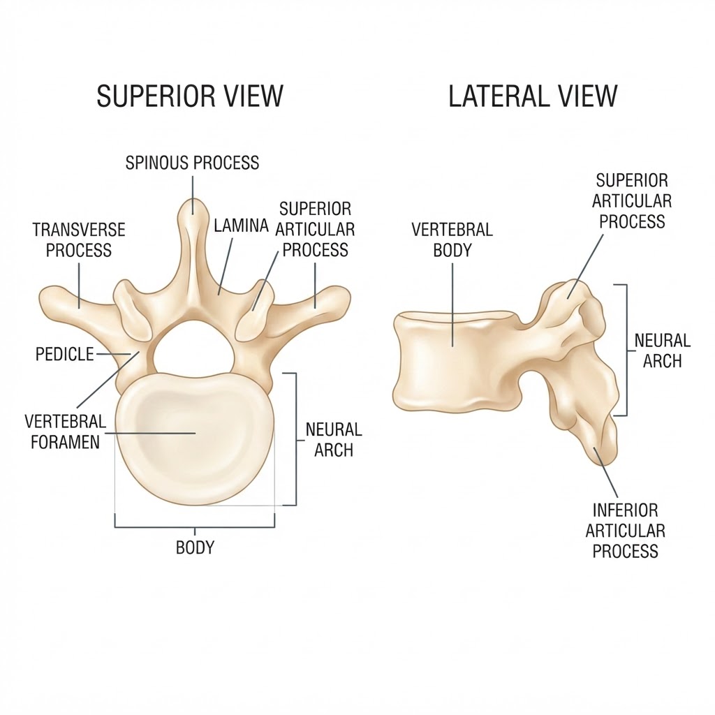
A labeled anatomical reference of a typical vertebra showing the vertebral body, neural arch, and key processes. Reviewer-rated 10/10 for anatomical accuracy.
Key Structures (Superior View)
- Body — large cylindrical anterior weight-bearing portion
- Vertebral foramen — central opening forming the vertebral canal when stacked
- Pedicle — short bony bridge connecting body to the arch
- Lamina — flat posterior part of the arch
- Transverse process — lateral projection for muscle/ligament attachment
- Spinous process — posterior midline projection (palpable down the back)
- Superior articular process — faces upward to form a facet joint
Key Structures (Lateral View)
- Vertebral body — anterior block
- Neural arch — posterior bony ring enclosing the foramen
- Superior & inferior articular processes — form the facet (intervertebral) joints
Clinical Note
- This image depicts a 'typical' vertebra; regional vertebrae differ — cervical have transverse foramina, thoracic have costal facets, lumbar have larger bodies
- Successive vertebral foramina form the vertebral canal, which houses the spinal cord
Review
- Verified anatomically correct against standard references (OpenStax)
- Reviewer score: 10/10
Source
- AI-generated full-color illustration, reviewer-verified
Like what you see?
Subscribe to unlock all 360 Study Cards and 15,000+ flashcards, track your progress, and study anywhere.